
Sensory Circuit Formation
Team Leader
Takeshi Imai (Ph.D .)
The mammalian central nervous system is composed of enormous numbers of neurons. How do these neurons establish their identity and form functional neuronal circuitries? To address this question, we are studying the mouse olfactory system as a model system. In the mouse olfactory system, odorants are detected by ~1,000 types of olfactory sensory neurons, each expressing a single type of odorant receptor. The olfactory bulb, the primary olfactory area of the brain, receives inputs from olfactory sensory neurons through 1,000 sets of glomeruli. These inputs are then processed in the olfactory bulb circuits and are then sent out to the olfactory cortex. These complex neuronal circuits emerge in an autonomous fashion based on cell-cell interactions and neuronal activity after birth, rather than by strict genetic programs. Using the olfactory bulb as a model system, we are trying to understand simple rules behind the formation of complex neuronal circuitry.
Select References
Iwata R, et.al. Mechanosensory-Based Phase Coding of Odor Identity in the Olfactory Bulb. Neuron 96, 1-14 (2017). doi:10.1016/j.neuron.2017.11.008
Murai A, et al. Distorted coarse axon targeting and reduced dendrite connectivity underlie dysosmia after olfactory axon injury. eNeuro 3, e0242-16.2016 (2016). doi:10.1523/ENEURO.0242-16.2016
Ke MT, et al. Super-resolution mapping of neuronal circuitry with an index-optimized clearing agent. Cell Rep 14, 2718–2732 (2016). doi:10.1016/j.celrep.2016.02.057
Imai T. Construction of functional neuronal circuitry in the olfactory bulb. Semin Cell Dev Biol 35, 180–188 (2014). doi:10.1016/j.semcdb.2014.07.012
Nakashima A, et al. Agonist-independent GPCR activity regulates anterior-posterior targeting of olfactory sensory neurons. Cell 154, 1314–1325 (2013). doi:10.1016/j.cell.2013.08.033
Ke MT, et al. SeeDB: a simple and morphology-preserving optical clearing agent for neuronal circuit reconstruction. Nat Neurosci 16, 1154–1161 (2013). doi:10.1038/nn.3447
Q:What made you want to do your current job? (or triggered your interest in science)
A:…
Q:What would you ultimately like to discover through your research?
A:…
Q:What is the greatest appeal of research?
A:…
Q:Do you have a policy, philosophy or motto that you apply to your research?
A:…
Q:If you were to characterize your lab in one word, what would if be?
A:…
Q:Scientist you respect (admire)
A:…
Q:Message for aspiring scientists.
A:…
Q:What interests or hobbies do you have outside of research?
A:…
Lab Homepage
imai[at]cdb.riken.jp
Recruit
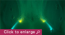
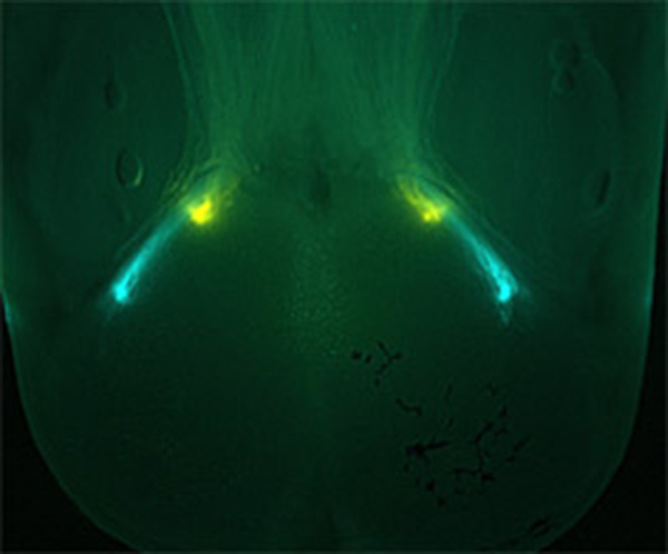
- Axonal projection of olfactory sensory neurons to the olfactory bulb. Cyan, olfactory sensory neurons expressing I7-CFP; Yellow, olfactory sensory neurons expressing I7-dnPKA-YFP.

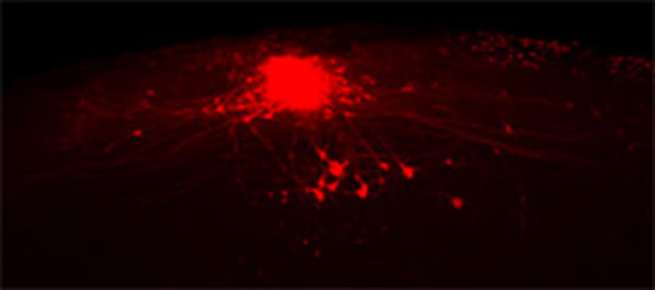
- Mitral and tufted cells in the mouse olfactory bulb labeled with TMR-dextran. Each mitral/tufted cell extends a single primary dendrite to a single glomerulus, where it receives inputs from a single specific type of olfactory sensory neurons.
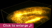
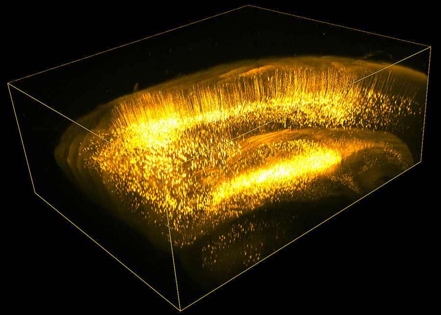
- Adult Thy1-YFP-H mouse brain was cleared with an optical clearing agent SeeDB and imaged using two-photon microscopy. 3D rendering image of a volume of 4mm x 5mm x 2mm, encompassing cerebral cortex and hippocampus, is shown.
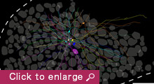
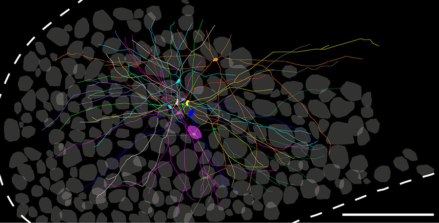
- Tracing of sister mitral cells associated with a common glomerulus in the olfactory bulb. Neurons connecting to a single glomerulus was labeled by neuronal tracer and the sample was cleared with SeeDB.

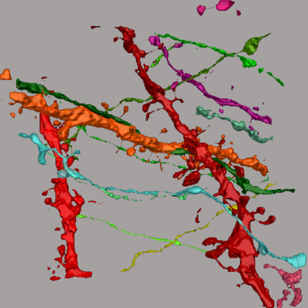
- Super-resolution mapping of neuronal circuitry. Brain slices of a Thy1-YFP-H transgenic mouse were cleared with SeeDB2, imaged with super-resolution microscopy, and reconstructed in 3D.
© RIKEN CENTER FOR DEVELOPMENTAL BIOLOGY All Rights Reserved.
![CDB [RIKEN CENTER FOR DEVELOPMENTAL BIOLOGY]](http://www.cdb.riken.jp/en/wp-content/themes/cdb_en/images/common/fLogo2.png)

